14 December, 2013
Capturing the Power of Digital Photography and the Precision of Laser Measurement to Advance Clinical Management
University of Maryland Medical Center, Baltimore, Maryland
Joan Lerner Selekof BSN, RN, CWOCN, Jeanette Massabni MS, RN, AGNP C, WOCN, Gwendolyn Williams BS, RN, CWON, Keisha McElveen MS, RN, CWON, Bret Anderson BA, RN, CWOCN, Julianna Sapp BSN, RN, CWOCN
Poster, SAWC Spring 2014
Introduction
Accurate wound management and valid tracking of healing trends is vital for informed decision making and documentation. The global cost of wound care is increasing, leading institutions to seek more efficient and accurate ways to improve wound assessment.
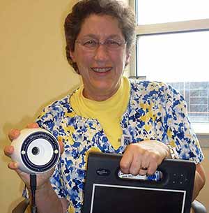
Joan Selekof, BSN RN CWOCN, Manager of the Center’s WOCN team, is pictured with a point of care wound surveillance device.
University of Maryland Medical System is rising to the challenges of managing this growing population of patients with wounds by integrating an electronic wound imaging system featuring high resolution digital photography with embedded laser measurement. This measurement and documentation is then captured directly into each patient’s electronic medical record.
- This system* provides objective data which accurately measures progress of wound healing (progression or deterioration), enhances communication between health care practitioners and patients.
- This system* also provides feedback to support staff education.
Method
This new technology, which was selected by the WOC Nurses and an interdisciplinary team from within the University of Maryland Medical System, has been initially used for pressure ulcer assessment and documentation. It provides a more efficient and accurate method to improve and communicate wound assessment, objective staging, healing trends and documentation. The three-dimensional laser triangulation-imaging device accounts for the curvature of the human body and three-dimensional nature of wounds to achieve highly accurate measurements. In addition to producing a high quality resolution image of the wound, software in the system* is integrated within the EMR and is accessible to authorized providers across the UMMS network using a secure centralized database.
Goals for System Implementation
- Implement a wound management system based on standardized documentation.
- Implement a wound management system across University of Maryland Medical System.
- Utilize the system* to track outcomes for patients whose wound records can follow the patient from hospital to hospital.
- Participate in translational research for wound management.
Implementation
- The implementation team included: IT project manger, CMO, surgeon and medical physician, WOC Nurses, members from the finance department, system analysts, risk management and the integration team.
- The interdisciplinary team decided to capture the wound image on admission, weekly, upon discharge and at the discretion of the practitioner.
- The WOC Nurse or the practitioner places the order in the EMR for wound imaging.
- The fundamental strength of this system* is that it accurately removes variances in measurement across users of the system* providing good intra-and inter-user reliability. Studies have shown this system’s* measurements are more accurate than both acetate tracings and digital planimetry from photos captured by standard digital cameras and smart phones.
- This technology tracks patient care across all venues of care and demonstrates improved clinical outcomes.
Results
Since its implementation by the WOC Nurses at University of Maryland Medical Center in May 2013, this new electronic wound imaging approach has provided a more efficient and accurate way to improve and communicate wound assessment, supporting objective staging, accurate healing trends and consistent documentation.
- Patient assessment reports generated by the system* into the EMR contain the progression of the wound over the patient’s hospitalization. History of the wound is available at any time within the electronic medical record.
- Case managers and other disciplines are now able to communicate accurate wound care documentation with imaging to home health agencies, acute care and long term care facilities upon patient’s discharge.
- Provides objective data which accurately measures progression or deterioration of wounds.
- Enhances communication between health care practitioners, patients, families and staff.
- Facilitates staff education on pressure ulcer prevention and wound assessment by presenting wound images in interdisciplinary rounds and “Huddles.”
- It is intended that this system* will support clinical trials and research initiatives, track the effectiveness of adjunctive therapies.
Discussion
With the ongoing challenges of health care reform, we must improve our clinical documentation. This technology was implemented initially to document pressure ulcers on admission and HAPU. The WOC nurses have since expanded their practice to include imaging any type of wounds. By embracing a new electronic wound imaging system*, University of Maryland Medical Center has improved communication between practitioners across the continuum. By providing accurate, objective imaging and a valid graph to track healing trends, we have improved patient care outcomes because we now have high quality, timely information on which to base our clinical decisions, intervene promptly, and determine best practice.
Maryland Medical Center case study – PDF version
References
- Kieser DC, Hammond C.Leading Wound Care Technology: The Aranz Medical Silhouette®. Adv Skin Wound Care 2011;24:68-70
- Bradshaw l, Gergar M, Hilko G. Collaboration in Wound Photography Competency Development: A Unique Approach. Adv Skin Wound Care 2011; 24:85-92
- Romanelli M, Rogers LC, Hammond CE, Nixon MA. Clinical evaluation of a wound measurement and documentation system. Wounds 2008;20:258-64
* Silhouette® Wound Assessment and Information System (ARANZ Medical Ltd) aranzmedical.wpengine.com


